Figure 2. Proliferative vitreoretinopathy epiretinal membranes. Immunohistochemical staining of α-smooth muscle actin showing immunoreactivity
in spindle-shaped myofibroblasts (arrows; A) and (B; original magnification 100×). Immunohistochemical staining of high-mobility group box −1 (HMGB1). Low-power (C; original magnification 40×) and high-power (D; original magnification 100×) showing spindle-shaped cells expressing cytoplasmic immunoreactivity to HMGB1 (arrows). Immunohistochemical
staining of the receptor for advanced glycation end products (RAGE) showing spindle-shaped cells expressing cytoplasmic immunoreactivity
to RAGE (arrows; E; original magnification 40×). Immunohistochemical staining of osteopontin showing strong cytoplasmic immunoreactivity in
spindle-shaped cells (arrows; F; original magnification 40×). Immunohistochemical staining of early growth response-1 showing spindle-shaped cells expressing
cytoplasmic immunoreactivity (arrows; G; original magnification 40×) and cells expressing nuclear immunoreactivity (arrows; H; original magnification 100×).
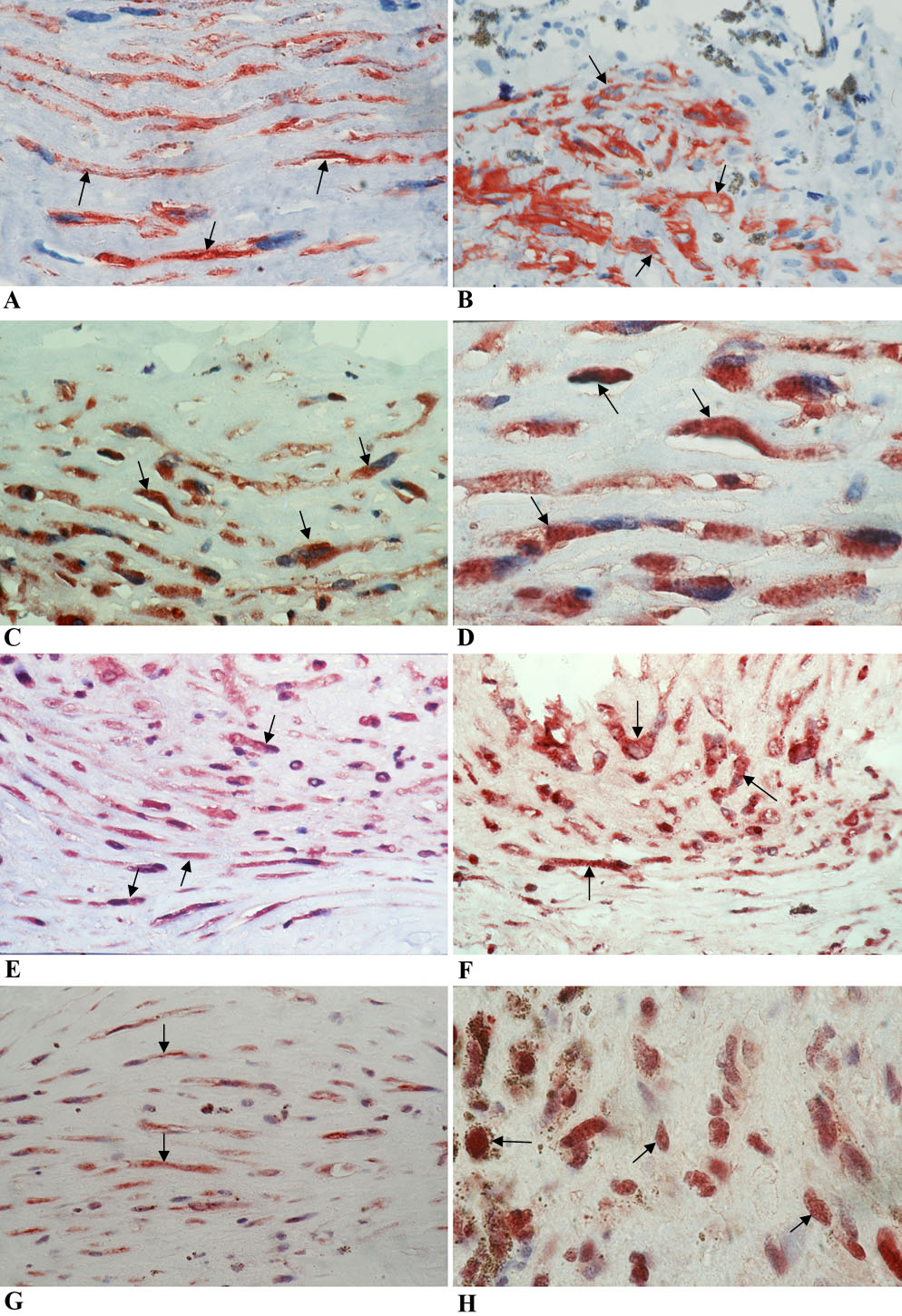
 Figure 2 of
Abu El-Asrar, Mol Vis 2011; 17:508-518.
Figure 2 of
Abu El-Asrar, Mol Vis 2011; 17:508-518.